 FMRI Studies
Click here for special information for MRI sessions for children.
FMRI stands for Functional Magnetic Resonance Imaging.
MRI is a painless imaging technology with no known harmful effects. It is used to gather information about the structure and functioning of the brain. MRI systems create a map of the tissues of the brain based on their magnetic properties. The brain’s changing blood flow and blood oxygenation allow researchers to use MRI to find which part of the brain is working during different tasks.
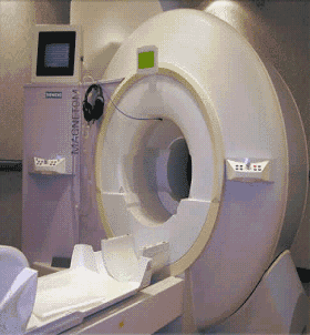
In 2012, the Lewis Center took delivery of its latest upgrade, a Siemens MAGNETOM Skyra 3T full-body MRI system. Along with many technological improvements, this system has a 70 cm bore, which will permit imaging of any part of the body and improve participant comfort. See the University’s Oregon Research press release for more information and photos of the installation.
 Only the head and neck are imaged with this research MRI system. A few people aren’t comfortable having their head still and in the dark enclosure. We do not use MRI if participants are not comfortable.
The University of Oregon’s MRI system is located in the Lewis Center for Neuroimaging.
Click here for How to prepare at home.
The purpose of these studies is to use magnetic resonance imaging (MRI) to study normal brain function as well as changes in brain function with changes in sensory, cognitive and language experience.
Procedures:
Pre-screening tests or questionnaires and consent forms are completed.
In order to become acclimated to the MRI environment an MRI simulator is provided which mimics what will be experienced in the actual scanner performing the same or similar tasks.
During these visits researchers give tests of memory, language or perception, and then take pictures both of the brain’s structure (standard MRI) as well as blood flow in the brain (functional MRI). These tests can involve finger tapping exercises, observing images through a mirror, listening to auditory stimuli, or solving an arithmetic problem and responding in accordance with instructions.
 All testing is carried out within the magnet and is conducted by a trained technician or research investigator. Specific information is given about the tasks and the duration of the study prior to being positioned within the MRI unit.